



An ECG and an echocardiogram (echo) are two commonly used tests for evaluating the heart, but they provide different types of information. An ECG records the electrical activity and rhythm of the heart, while an echocardiogram uses ultrasound to create images of the heart and assess its structure and function.
So, what is the difference between ECG and echo?
An ECG can help doctors assess heart rate, rhythm and certain electrical abnormalities. An echocardiogram can provide information about the heart chambers, valves, muscle movement, pumping function and blood flow.
Neither test is universally “better.” A cardiologist may recommend an ECG, an echo or both depending on your symptoms, medical history, examination and the clinical question that needs to be answered.
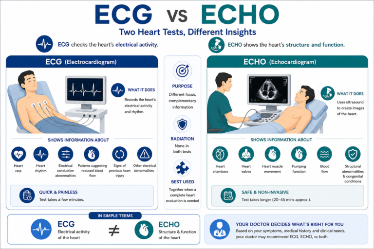
ECG stands for electrocardiogram.
An ECG is a test that records the electrical signals produced by the heart. The heart’s electrical system controls the timing and rhythm of each heartbeat. An ECG captures these electrical signals through electrodes placed on the skin.
A standard ECG may help a healthcare professional evaluate:
An ECG does not create an image of the heart. Instead, it produces a tracing that represents the heart’s electrical activity.
An echocardiogram, commonly called an echo, is an ultrasound-based test used to create images of the heart. A standard transthoracic echocardiogram uses an ultrasound probe placed against the chest. The sound waves create images that allow the healthcare team to assess different parts of the heart.
An echocardiogram can provide information about:
Unlike an ECG, an echocardiogram provides visual information about the heart’s structure and mechanical function.
| Feature | ECG | Echocardiogram |
|---|---|---|
| Full name | Electrocardiogram | Echocardiogram |
| Main purpose | Records electrical activity | Creates images of the heart |
| How it works | Electrodes record electrical signals | Ultrasound waves create heart images |
| Main information | Heart rate, rhythm and electrical patterns | Structure, movement, valves and pumping function |
| Produces an image of the heart? | No | Yes |
| Assess heart rhythm? | Yes | Not its primary purpose |
| Assess heart valves? | Not directly | Yes |
| Assess the pumping function? | Not directly | Yes |
| Uses ionizing radiation? | No | No |
| Usually invasive? | No | Standard transthoracic echo is non-invasive |
| Common use | Electrical/rhythm assessment | Structural and functional assessment |
If a doctor wants to assess the heart’s electrical activity or rhythm, an ECG may be particularly useful.If the doctor needs to evaluate the heart’s structure, valves, muscle movement or pumping function, an echocardiogram may provide more relevant information.
In some situations, a doctor may recommend both tests because they provide different types of information.The appropriate test depends on the patient’s symptoms, medical history, examination findings and suspected condition.
An ECG can provide information about the heart’s electrical activity. Depending on the clinical situation, it may help identify:
However, an ECG cannot detect every type of heart problem.A normal ECG does not necessarily mean that every aspect of the heart is completely normal. Additional testing may sometimes be required based on symptoms and clinical findings.
An echocardiogram provides images that can help assess the heart’s structure and function.
It may help evaluate:
The size and structure of the heart chambers can be assessed.
An echo can help evaluate the appearance and movement of heart valves and identify certain valve abnormalities.
The movement and thickness of the heart muscle can be assessed.
An echocardiogram can help evaluate how effectively the heart pumps blood.
Doppler techniques can be used during echocardiography to assess blood flow through the heart and valves.
An echo can help identify certain structural problems and some congenital abnormalities.
Sometimes, yes. An ECG and an echocardiogram provide different information, so a cardiologist may recommend both when a more complete evaluation is needed.
For example, a patient may have symptoms that require assessment of both:
However, not everyone with a heart-related symptom needs both tests.Your doctor will determine which investigations are appropriate based on your individual circumstances.
An ECG may be recommended in situations such as:
The exact reason for performing an ECG depends on the patient’s symptoms and clinical history.
A doctor may recommend an echocardiogram when they need information about the structure or function of the heart.
Possible reasons include:
An echocardiogram may also be used to monitor how a known heart condition changes over time.
A standard ECG is generally painless. Small electrodes are placed on the skin of the chest and sometimes the arms and legs. The electrodes detect electrical signals from the heart.
You may feel mild discomfort when electrodes are removed, particularly if they adhere firmly to the skin, but the test itself does not send electricity into your body.
In your 30s, work and family responsibilities can make healthy habits harder to maintain. This is a good time to pay attention to:
Do not assume that cardiovascular problems only become relevant after 40.
A standard transthoracic echocardiogram is generally painless and non-invasive.The healthcare professional places an ultrasound probe against the chest using a special gel.
Some pressure may be applied to obtain clearer images, which can occasionally feel slightly uncomfortable, particularly if the chest area is already tender. An echocardiogram does not use ionizing radiation.
A standard resting ECG is usually a relatively quick test. The actual recording generally takes only a few minutes, although the overall appointment may take longer because of preparation, electrode placement and review. The exact duration can vary depending on the type of ECG and the clinical setting.
A standard echocardiogram generally takes longer than a resting ECG because multiple images and measurements of the heart are obtained. The duration can vary depending on the type of echocardiogram, the patient’s condition and the complexity of the examination.
Your healthcare team can provide more specific information about the expected duration of your appointment.
No. An ECG is an important cardiac test, but it evaluates primarily the heart’s electrical activity. Some structural or functional problems may require imaging such as echocardiography or other investigations.
For example, an ECG alone cannot provide the same information about heart valves, chamber structure or pumping function that an echocardiogram can provide. This is why doctors may use several tests together when clinically appropriate.
A standard echocardiogram primarily evaluates the heart’s structure and function. It is not the same as a test specifically designed to visualize coronary artery blockages.
Depending on the clinical situation, doctors may recommend other investigations to evaluate coronary artery disease.
A stress echocardiogram, for example, evaluates the heart’s response to stress and can provide additional information, but it should not be assumed to be interchangeable with tests that directly visualize coronary arteries.
Your cardiologist can determine which test is appropriate based on your symptoms and risk factors.
ECG and echocardiography are only two of the tests that may be used in cardiac evaluation. Depending on the clinical situation, a cardiologist may also consider:
Each test answers different clinical questions. The goal is not to undergo as many tests as possible. The goal is to use the right test for the clinical question.
You should consider discussing your symptoms with a healthcare professional if you experience:
If a doctor has already recommended an ECG, echocardiogram or another cardiac investigation, follow their advice and ask what the test is intended to evaluate.
Seek Urgent Medical Attention for Concerning Symptoms
Sudden or severe chest discomfort, severe breathlessness, fainting or symptoms that may indicate a heart attack require urgent medical attention.
Do not delay emergency evaluation while waiting to schedule a routine cardiac test.
If you have been advised to undergo cardiac testing or have symptoms that require evaluation, it is important to have the tests performed in an appropriate clinical setting and interpreted in the context of your overall health.
Smt S R Mehta & Sir K P Cardiac Institute provides specialised cardiac evaluation and care in Mumbai.
ECG and echocardiography are complementary heart tests, not interchangeable tests. An ECG evaluates the electrical activity and rhythm of the heart, while an echocardiogram uses ultrasound to assess the heart’s structure and function.
Your doctor may recommend an ECG, an echo or both depending on your symptoms, medical history and examination findings.
If you have symptoms such as chest discomfort, unexplained breathlessness, palpitations, dizziness or fainting, do not rely on an online comparison to decide which test you need. A qualified healthcare professional can determine the appropriate evaluation.
For patients seeking specialised cardiac evaluation and care in Mumbai, Smt S R Mehta & Sir K P Cardiac Institute provides cardiac services and diagnostic evaluation.
Written by: Smt S R Mehta & Sir K P Cardiac Institute
Medically Reviewed by:Smt S R Mehta & Sir K P Cardiac Institute
Medical Review: This article has been reviewed for medical accuracy by a qualified cardiac specialist. It is intended for general education and does not replace an individual medical consultation or diagnosis.
S.R. Mehta & Sir K.P. Cardiac Institute, Mumbai’s premier facility noted for excellence in heart health services, provides the greatest cardiac hospital experience.
WhatsApp us