


Medical diagnostic imaging refers to non-invasive technologies such as X-rays, MRI, CT scans, and Ultrasound that allow physicians to view internal organs, bones, and tissues without surgery. While X-rays and CT scans utilize controlled ionizing radiation for fast bone and trauma evaluation, MRI (magnetic fields) and Ultrasound (sound waves) are 100% radiation-free, making them ideal for detailed soft tissue, brain, and cardiac or prenatal assessments.
Medical imaging has transformed modern healthcare by allowing doctors to diagnose diseases quickly, accurately, and often before symptoms become severe. Whether you’re experiencing persistent pain, recovering from an injury, or undergoing a routine health check-up, the right diagnostic imaging test can help identify the underlying cause and guide the most effective treatment.
Today, several imaging techniques are available, each designed for a specific purpose. Some are best for examining bones, while others provide highly detailed images of soft tissues, internal organs, or blood vessels.At Smt. S.R. Mehta & Sir K.P. Cardiac Institute, advanced diagnostic imaging services support early disease detection, enabling specialists to diagnose conditions accurately and recommend timely treatment.
In this guide, we’ll explain the 7 most common types of diagnostic imaging X-ray, MRI, CT scan, and Ultrasound; their uses, benefits, differences, and when your doctor may recommend each one.
Choosing the right diagnostic imaging test is essential for accurate diagnosis and effective treatment. In this guide, you’ll learn how X-rays, MRI scans, CT scans, and Ultrasound work, when each test is recommended, their benefits, safety considerations, and how healthcare professionals select the most appropriate imaging method based on your symptoms and medical condition.
Diagnostic imaging refers to a group of medical tests that create images of structures inside the body without surgery. These images help doctors detect injuries, infections, tumors, fractures, heart disease, organ abnormalities, and many other medical conditions.
Modern diagnostic imaging plays an essential role in:
Because each imaging technique serves a different purpose, your doctor will recommend the most appropriate test based on your symptoms and medical history.
Did You Know?
Many serious conditions, including certain cancers, heart diseases, strokes, and internal injuries, can be detected at an early stage through diagnostic imaging, often before noticeable symptoms develop.
Diagnostic imaging provides doctors with detailed information that cannot be obtained through a physical examination alone. It supports faster diagnosis, helps determine the severity of a condition, and guides treatment planning.
Healthcare professionals rely on imaging tests to:
Expert Tip:
Your doctor selects an imaging test based on your symptoms, medical history, age, and the body part being examined. The most advanced scan is not always the most appropriate one choosing the right test improves diagnostic accuracy while avoiding unnecessary procedures.
Imaging Type | Primary Source | Radiation? | Primary Focus |
X-Ray | X-Ray Beams | Low | Bone & Density |
MRI | Magnetic Fields | None | Soft Tissue & Nerves |
CT Scan | 360° X-Rays | Moderate | 3D Cross-Sections |
Ultrasound | Sound Waves | None | Real-Time Motion |
PET Scan | Radioactive Tracer | Moderate | Cellular Metabolism |
Mammography | Low-Dose X-Ray | Very Low | Breast Tissue |
Fluoroscopy | Continuous X-Ray | Moderate | Live Video / Motion |
X-rays are the foundation of medical diagnostics. They pass a minimal, safe dose of targeted ionizing radiation through the body. Because denser structures like bones absorb more radiation, they appear bright white on the film, while soft tissues appear in shades of gray.
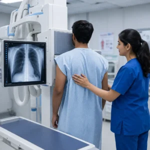
Safety Note: Modern digital X-rays use up to 80% less radiation than traditional film X-rays, making them extremely safe for routine diagnostic use.
Magnetic Resonance Imaging (MRI) uses powerful radio waves and a magnetic field to manipulate hydrogen atoms in the body’s cells. A computer measures the energy released by these atoms to construct high-contrast images of organs and soft tissues.
Because MRI involves zero radiation, it is safe for repeated scans when monitoring chronic conditions.
A CT (or CAT) scan takes X-ray imaging a step further by rotating 360 degrees around the patient’s body. A computer compiles these slice-by-slice X-ray images into detailed 3D cross-sectional models of bones, blood vessels, and internal organs simultaneously.
Ultrasound uses high-frequency sound waves transmitted through a hand-held probe (transducer) coated with sound-conducting gel. As sound waves bounce off internal structures, real-time visual images are displayed on a monitor.
Unlike structural scans (X-ray or CT), a PET Scan evaluates how your cells and organs are functioning at a biochemical level. Before the scan, a small, safe amount of a radioactive tracer (such as fluorodeoxyglucose or FDG) is injected into the bloodstream. Cancerous or hyperactive cells absorb this tracer at a higher rate, lighting up on the scan.
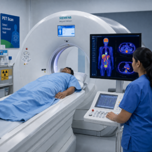
Key Distinction: CT and MRI show what a disease looks like structurally, while a PET scan shows how cellular tissue is functioning. Modern systems often combine these into PET-CT scans for pinpoint accuracy.
A Mammogram is a specialized, low-dose X-ray system designed specifically to examine breast tissue. It compresses the breast briefly between two plates to spread out the tissue, allowing low-dose radiation to detect abnormal growths or microcalcifications long before they can be felt during a physical exam.
If a standard X-ray is like a photograph, Fluoroscopy is like a movie. It passes a continuous X-ray beam through the body to display a live, moving image on a monitor. Patients often ingest or are injected with a contrast medium (like barium or dye) so physicians can watch internal organs or fluids function in real time.
The appropriate imaging test depends on your symptoms and the area of the body being examined.
Condition | Recommended Imaging |
Bone fracture | X-Ray |
Brain disorder | MRI |
Sports injury | MRI |
Stroke | CT Scan |
Internal bleeding | CT Scan |
Pregnancy | Ultrasound |
Gallstones | Ultrasound |
Kidney stones | CT Scan or Ultrasound |
Chest infection | X-Ray |
Preparation varies depending on the imaging procedure. Following your doctor’s instructions helps produce clear images and reduces the need for repeat examinations.
Preparation depends on the area being examined.
Expert Tip: Following preparation instructions carefully can improve image quality and reduce the need for repeat imaging.
Yes. Diagnostic imaging procedures are generally safe when performed under medical supervision.
Healthcare providers carefully evaluate the benefits and risks before recommending any imaging test.
Diagnostic imaging should always be performed based on a doctor’s clinical evaluation. Seek medical attention if you experience:
Early medical evaluation can help identify serious health conditions before they become more advanced.
Choosing an accredited imaging facility with experienced radiologists ensures you receive accurate reports and timely interventions.
Located centrally in Sion, Smt. S.R. Mehta & Sir K.P. Cardiac Institute provides complete, round-the-clock radiology and diagnostic imaging services:
If your physician has ordered an imaging scan or if you require an expert second opinion, visit our central facility in Sion, Mumbai.
Book an Imaging Appointment or Consultation at SRM Cardiac Institute
This article is based on guidance and educational information from internationally recognized healthcare organizations, including:
These organizations provide evidence-based recommendations on the safe and effective use of diagnostic imaging in clinical practice.
Diagnostic imaging has become an essential part of modern healthcare, enabling doctors to diagnose medical conditions with greater accuracy and confidence. Whether evaluating a suspected fracture, investigating persistent pain, assessing internal organs, or monitoring a chronic condition, imaging tests provide valuable information that supports timely and effective treatment.
Understanding the differences between X-ray, MRI, CT scan, and ultrasound helps patients make informed decisions and better understand why a specific imaging test has been recommended. Each technique has unique strengths, and selecting the appropriate test is based on your symptoms, medical history, and clinical findings.
If you’re looking for reliable and advanced diagnostic imaging services in Mumbai, Smt. S.R. Mehta & Sir K.P. Cardiac Institute offers state-of-the-art imaging technology, experienced radiologists, and comprehensive diagnostic care under one roof. Early diagnosis can make a significant difference in treatment outcomes and schedule your consultation today for accurate evaluation and timely care.
Accurate diagnosis begins with high-quality imaging. At Smt. S.R. Mehta & Sir K.P. Cardiac Institute patients benefit from advanced diagnostic technology and experienced specialists who ensure precise reporting and timely diagnosis.
Our diagnostic imaging services include:
Whether you need routine imaging or advanced diagnostic evaluation, our multidisciplinary team is committed to delivering reliable results and quality patient care.
If your doctor has recommended an imaging test, choosing a trusted diagnostic center can make a meaningful difference in the accuracy of your diagnosis.
At Smt. S.R. Mehta & Sir K.P. Cardiac Institute, our experienced radiologists and advanced imaging technology support early detection and informed treatment decisions across a wide range of medical specialties.
Schedule your diagnostic imaging appointment today and take the next step toward timely, accurate healthcare.
Disclaimer: This article is intended for educational and informational purposes only. It should not be considered a substitute for professional medical advice, diagnosis, or treatment. Always consult a qualified healthcare professional regarding your symptoms, medical condition, or recommended diagnostic imaging procedure.
Smt. S.R. Mehta & Sir K.P. Cardiac Institute
Specialization: Diagnostic & Interventional Radiology
Content Reviewed & Published by
Smt. S.R. Mehta & Sir K.P. Cardiac Institute
Our healthcare content is developed to provide accurate, patient-friendly information based on current medical knowledge and is medically reviewed by experienced specialists before publication.
You can also explore: